Scalp Acupuncture for Stroke Rehabilitation
Scalp Acupuncture, also known as Neuroacupuncture, is a contemporary acupuncture method. It is based on the modern the modern knowledge of biomedical anatomy, neurology and physiology of the brain combined with the Traditional Chinese Medicine theory and acupuncture needle techniques.
- Scalp acupuncture
- Stroke
- Scalp Acupuncture for Stroke rehabilitation and Mental Health
- Contraindications to scalp acupuncture
- Localization and Indications of Head Stimulation Areas
- Stimulation zone positioning and indications
- Acupoint selection method
Scalp acupuncture
Scalp acupuncture, also known as Neuroacupuncture, is a micro-system acupuncture technique which stimulate the areas or points on the scalp to treating brain associated diseases, neurological and psychological conditions, et al. It is one of the modern acupuncture therapies based on the traditional Chinese medicine (TCM) theory, acupuncture needling techniques combined with neurology and physiology of the brain.
Acupuncture can help stroke survivors improve mobility, sensation, speech, increase muscle strength, and promote overall rehabilitation.
Stroke
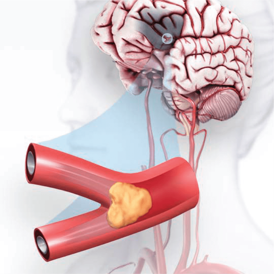 A stroke is a medical condition, sometimes called a brain attack, in which poor blood supply to part of the brain or a blood vessel in the brain bursts causes cell death.(The picture is from America Stroke Association.)
Three types of stroke:
- Ischemic stroke:
a blood clot obstructing the flow of blood to the brain
- Hemorrhagic stroke:
a blood vessel rupturing and preventing blood flow to the brain
- Transient ischemic attack(TIA):
“mini stroke”, is caused by a temporary clot
Scalp Acupuncture for Stroke rehabilitation and Mental Health
- Scalp acupuncture is a therapy that treats diseases by stimulating the points or areas on the scalp to treating brain related diseases, neurological and psychological conditions, et al.
- Based one traditional Chinese medicine theory, the scalp acupuncture is a micro-system acupuncture combined acupuncture needling techniques with neurological knowledge.
- Scalp acupuncture was proposed as early as the 1950s , but it was really promoted clinically after the 1970s . Through the treatment of a large number of patients, it has been proved that scalp acupuncture is not only simple and safe, but also has unique effects on various diseases caused by the brain.
Contraindications to scalp acupuncture
- Patients with cerebral hemorrhage who are in a coma or whose condition is not stable should not use it.
Localization and Indications of Head Stimulation Areas
Positioning basis
- There are two commonly used ones: one is to select relevant meridians and points on the head for treatment according to the theory of viscera and meridians.
- The second is to divide the corresponding stimulation areas on the scalp for acupuncture according to the functional positioning of the cerebral cortex. The second is mainly introduced below.
- It should be noted that the projection position of the brain gyrus on the cranial surface and the currently established stimulation site can only be said to be basically corresponding. From the examination of CT photos, there are a certain number of individual differences. When delineating the stimulation line, the skull should be properly considered. shape.
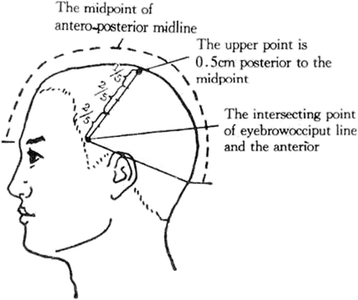
Positioning line
Two standard positioning lines demarcating the stimulation area.
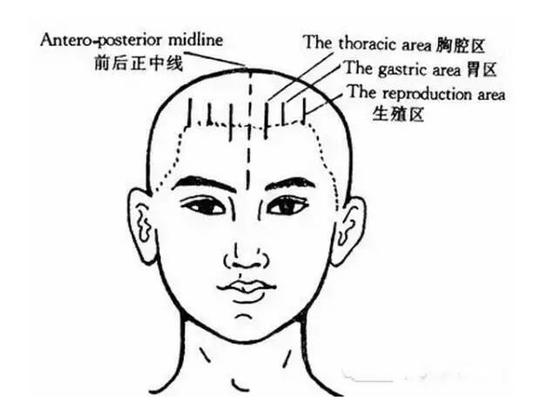
- Anteroposterior midline: It is a line from the midpoint between the two brows (the point in front of the center) to the lower edge of the tip of the extraoccipital tuberosity (the point behind the midline) passing through the top of the head.
- Brow-occipital line: It is a line connecting the upper border of the eyebrow midpoint and the tip of the extraoccipital tuberosity on the side of the head.
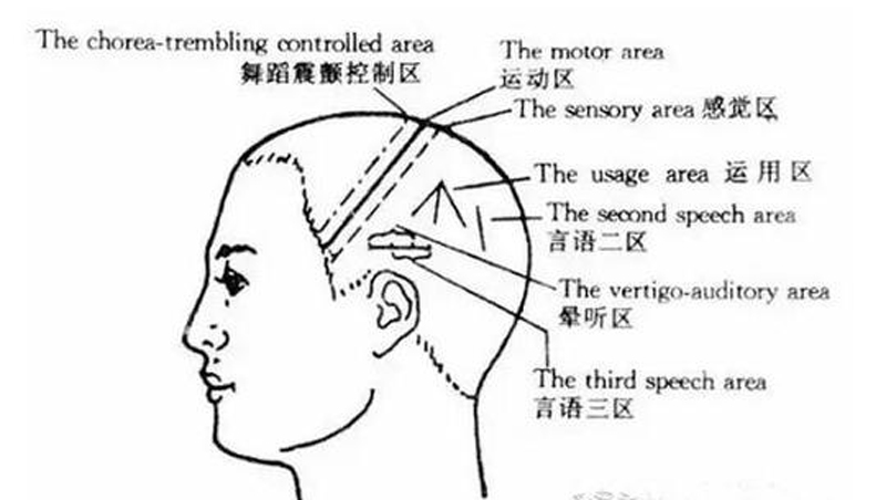
Stimulation zone positioning and indications
Sports area
Location: The upper point is 0.5 cm behind the midpoint of the front and rear midlines; the lower point is at the intersection of the brow-occipital line and the front edge of the temple hairline. If the sideburns are not obvious, a vertical line can be drawn upward from the midpoint of the zygomatic arch, and the intersection of this line and the brow-occipital line is moved forward by 0.5 cm as the lower point of the motor zone. The line connecting the upper and lower points is the operating area. The sports area can be divided into upper, middle and lower parts.
-
Upper part: the upper 1/5 of the upper motor area, which is the lower limb and trunk motor area.
-
Middle part: It is the middle 2/5 of the motor area, which is the upper limb motor area.
-
Lower part: It is the lower 2/5 of the motor area, which is the facial motor area, also known as the speech area.
Attending
-
Upper part: paralysis of contralateral lower limbs and trunk.
-
Middle part: paralysis of contralateral upper limb.
-
Lower part: Contralateral central facial nerve paralysis, motor aphasia (partial or complete loss of language ability, but basically retains the ability to understand language), salivation, dysphonia.
Sensory zone
Location: The parallel line moving back 1.5 cm in the motor area is this area. The sensory area can be divided into upper, middle and lower parts.
-
Upper part: It is the upper 1/5 of the sensory area, which is the sensory area of the lower limbs, head, and trunk.
-
Middle part: It is the middle 2/5 of the sensory area, which is the upper limb sensory area.
-
Bottom: It is the lower 2/5 of the sensory area, which is the surface sensory area.
Indications:
-
Upper part: Contralateral lumbago and leg pain, numbness, paresthesia, back of the head, neck pain, dizziness, tinnitus.
-
Middle part: Pain, numbness and paresthesia in the contralateral upper limb.
-
Lower part: Contralateral facial numbness, migraine, temporomandibular head arthritis, etc.
The chorea-trembling controlled area
Location: Parallel line moving 1.5 cm forward in the motor zone.
Indications: chorea, parkinsonism, parkinsonism syndrome.
Dizziness area
Location: 1.5 cm straight up from the tip of the ear, draw a horizontal line 2 cm forward and 2 cm backward.
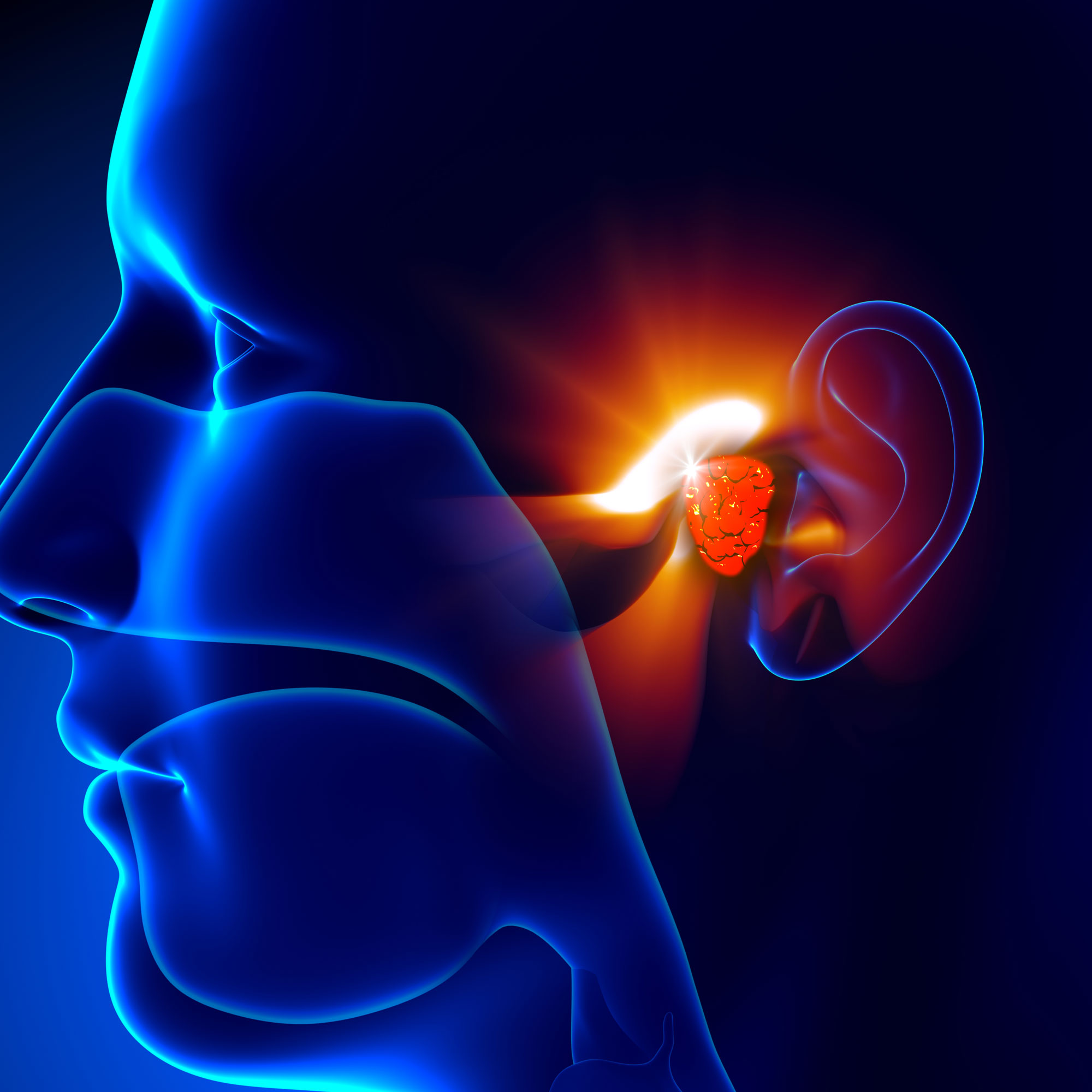
Indications: dizziness, tinnitus, hearing loss.
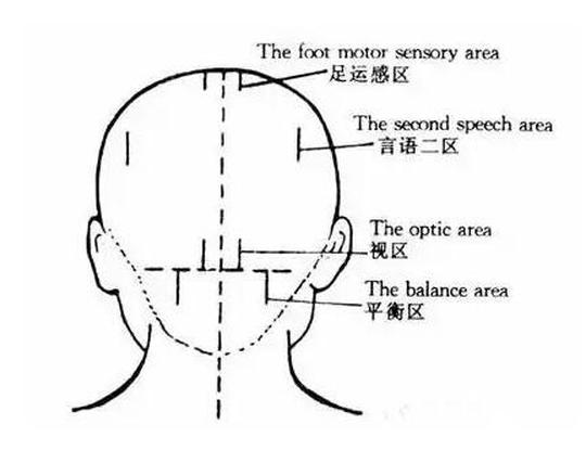
The second speech area
Location: Draw a straight line parallel to the anteroposterior midline from 2 cm behind the parietal tuberosity, and draw a 3 cm long straight line downward.
Indications: Nomenclature aphasia. (Also known as amnestic aphasia, the patient’s ability to call “name” is impaired. For example, the patient can’t call “chair”, but only “sitting”; when others call chair, he can understand.) The third speech area
Location: A horizontal line 4 cm long is drawn backward from the midpoint of the hearing fainting area.
Indications: sensory aphasia. (Patients have impaired ability to understand speech and often answer questions that are not asked.)
The usage area
Location: Draw a vertical line from the parietal tuberosity and two lines with an angle of 40 degrees to the line, both of which are 3 cm in length.
Indications: apraxia. (Also known as apraxia, the patient’s ability, muscle tone and basic movement are normal, but there is a technical ability obstacle, such as being unable to unbutton, pick up coins, etc.)
The foot motor sensory
Location: 1 cm away from the midpoint of the front and rear midlines, 1 cm to the left and right, and 3 cm long to the back, parallel to the midline.
Indications: Contralateral lower limb paralysis, pain, numbness, acute lumbar sprain, nocturia, cortical polyuria, uterine prolapse, etc.
The optic area
Location: On the horizontal line of the outer occipital tuberosity 1 cm away from the posterior point of the anterior-posterior midline, draw upward a 4-cm-long straight line parallel to the anterior-posterior midline.
Indications: cortical visual impairment.
The balance area
Location: On the horizontal line of the extraoccipital tuberosity 3.5 cm away from the posterior point of the anterior-posterior midline, draw a 4-cm long straight line parallel to the anterior-posterior midline downward.
Indications: ataxia caused by cerebellar disease, balance disorder, dizziness, limb numbness and paralysis caused by brain stem dysfunction.
The gastric area
Location: Starting from the pupil directly above the hairline, go upward and parallel to the anteroposterior midline with a length of 2 cm.
Indications: Stomach pain and upper abdominal discomfort caused by gastritis and gastric ulcer.
The thoracic area
Location: Between the stomach area and the front and rear midlines, draw a 2 cm long straight line above and below the hairline.
Indications: bronchial asthma, chest discomfort and other diseases.
The reproduction area
Location: Draw a 2 cm long straight line parallel to the anteroposterior midline upward from the forehead.
Indications: dysfunctional uterine bleeding, pelvic inflammatory disease, uterine prolapse, etc.
Acupoint selection method
- For unilateral limb disease, choose the contralateral stimulation area; for bilateral limb disease, choose the bilateral stimulation area; The relevant stimulated area cooperates with the treatment. For lower extremity paralysis, the lower extremity motor area can be combined with the foot movement sensory area.


Comments
Nothing yet.